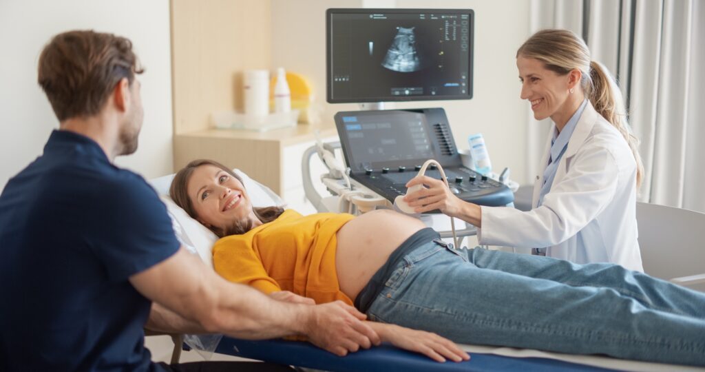
Ultrasound plays a key role in monitoring pregnancy health. Research shows that exams performed in specialized centers—under the supervision of MFM specialists—have higher detection rates for fetal, placental, and cervical abnormalities.
Types of Prenatal Ultrasound Studies
First Trimester Anatomy / (11–14 weeks)
This ultrasound evaluates early fetal anatomy, confirms dating. This measurement helps refine the risk assessment for chromosomal abnormalities and certain structural issues.
Nuchal Translucency (12–13+6 weeks)
Our office performs nuchal translucency (NT) ultrasounds between 12 weeks and 13 weeks 6 days of pregnancy. The NT scan is a specialized ultrasound that measures the small pocket of fluid at the back of the baby’s neck. This measurement helps assess the risk for certain chromosomal conditions, such as Down syndrome (Trisomy 21), as well as other genetic conditions and some structural abnormalities, including congenital heart defects.
While the NT scan is separate from a routine first-trimester ultrasound, it can only be performed during the first trimester, within the specific gestational age window noted above. When combined with maternal age and optional bloodwork, the NT scan provides valuable early information about your baby’s development.
Detailed Anatomy Ultrasound (18–22 weeks)
A comprehensive evaluation of fetal development and a primary screening exam for birth defects.
This study includes detailed assessment of:
- Brain and spine
- Heart
- Face
- Limbs
- Kidneys and abdomen
- Placenta
- Amniotic fluid
- Cervix
This exam also screens for placental problems and can identify risk factors for preterm birth.
Fetal Echocardiogram (>20 weeks)
Can be performed as early as 16 weeks if a heart abnormality is suspected.
A specialized study focused on the baby’s heart structure and function.
Common indications include:
- Maternal diabetes
- Family history of congenital heart disease
- Abnormalities detected on prior ultrasound
- Certain genetic conditions
- IVF Pregnancy
Growth Ultrasound (>20 weeks)
Evaluates fetal size, weight, and interval growth.
Growth ultrasounds are commonly ordered for:
- High-risk pregnancies
- Maternal medical conditions (e.g., hypertension, diabetes)
- Suspected fetal growth restriction
- Monitoring twins or higher-order multiples
Doppler Studies
Doppler ultrasound evaluates blood flow in key fetal and placental vessels.
Common indications include:
- Suspected fetal growth restriction
- Evaluation of placental insufficiency
- Monitoring pregnancies complicated by isoimmunization
- Assessing fetal anemia via middle cerebral artery Doppler
- Twin Gestations
Doppler findings help guide timing of delivery and fetal surveillance.
Biophysical Profile (BPP)
A BPP assesses fetal well-being by evaluating:
- Fetal movement
- Fetal breathing
- Muscle tone
- Amniotic fluid
When combined with fetal heart rate monitoring, it provides an overall assessment of fetal well-being.
Common indications include high-risk pregnancies, decreased fetal movement, maternal hypertension, diabetes, or growth concerns.
Transvaginal Ultrasound
A specialized technique used when abdominal imaging does not provide sufficient detail.
Common indications include evaluation of:
- Cervical length to assess preterm birth risk
- Placental and umbilical cord position (placenta previa or low-lying placenta)
- Uterus and ovaries (especially early in pregnancy)
- Early fetal development when the uterus is small or when fetal position limits visibility
This exam is safe, well-tolerated, and provides essential diagnostic information.